 |
So-called idiopathic scoliosis |  |
| SKOLIOZA / SCOLIOSIS | (14) Scoliosis in English |  POLISH VERSION POLISH VERSION |
Partner / książki / books: 
|
Biomechanical aetiology of the so-called idiopathic scoliosis (1995 – 2007). New clinical and radiological classification. Rules of new rehabilitation treatment and causal prophylactics Prof. Tomasz Karski MD PhD / x Head (1995-2009) of Chair and Department of Paediatric Orthopaedic and Rehabilitation Medical University of Lublin, University Paediatric Hospital, Chodźki Street 2 20-093 Lublin, Poland, tel./fax 081/741 56 53 x/ (2010) Vincent Pol University in Lublin / Poland E-mail: t.karski@neostrada.pl tkarski@dsk.lublin.pl www.ortopedia.karski.lublin.pl Key words
Abstract The article presents biomechanical etiology of the so-called idiopathic scoliosis [adolescent idiopathic scoliosis - AIS] (1995-2007). Development of scoliosis is connected with “gait” and habitual “standing at ease” on the right leg. This new classification of AIS describes: “S” shaped scoliosis in group I etiopathological group (epg), “C” shaped scoliosis – in group II/A epg and “S” shaped scoliosis – in group II/B epg and also “I” shaped scoliosis – in group III epg. These groupings were distinguished in the years 2001-2004 / 2006. In all children with so-called idiopathic scoliosis limited adduction of the right hip was found, or even abduction contracture of this hip, often connected with flexion and external rotation contracture. The contracture, or difference in adduction range (smaller in the right hip), is connected with the “syndrome of contractures” in newborns and babies described by many authors but thoroughly and very exactly by Prof. Hans Mau. The new classification clarifies the therapeutic approach to each etiopathological group of scoliosis and provides the possibility to introduce causative prophylaxis.
1. Introduction: Through many years the aetiology of the so-called idiopathic scoliosis (AIS) was unknown. Many researchers searched for “etiological factors” of scoliosis including genetic, hormonal factors, growth abnormalities, neuromuscular influences, disorders in bones, disorders in muscle and fibrous tissue, growth rate, left – right symmetry / asymmetry (and here directional asymmetry, anti-asymmetry, fluctuating asymmetry), anterior – posterior symmetry / asymmetry (and here directional asymmetry, anti-asymmetry, fluctuating asymmetry), asymmetry in growth of spinal cord and vertebra bodies and in “asymmetry concept” – arm length, facial structure, trunk, hand & foot preference, “reducing” asymmetry with age, nervous system lateralization, dermatoglyphics, developmental instability, “boy gait” versus “girl gait”, thoracic-spinal deformity primary as concept for idiopathic scoliosis – “multi-factorial” – “silent” concept, CNS, immature scoliotic vertebrae, circulating factor and plenty of other hypothetic influences (taken from Second Round EFG 6 Discussion / International Federated Body on Scoliosis Aetiology (IBSE) / Electronic Focus Group-6). Our first presentation on “biomechanical influences for development of the spine” was in 1995 in Hungary (Orthopaedic Congress in Szeged). The biomechanical aetiology of the so-called idiopathic scoliosis is based on asymmetry of movements of left and right hips and resulting asymmetry of loading during gait, leading to asymmetry of growth between left and right side of the body. This results over time as scoliosis. All children with so-called idiopathic scoliosis have the habit of permanently standing “at ease” only on the right leg. This is connected with “real abduction contracture” or otherwise limited / restricted adduction of the right hip often with co-existing flexion and external rotation contractures in comparison to the left hip with full movements. This hip asymmetry makes the right leg “stronger”, “more stable”, and consequently “more easy for standing on”. This asymmetry of movements is connected with the “syndrome of contractures” in newborns and babies (Originally in German - “Siebenersyndrom” – Prof. Hans Mau Tübingen / Germany).
2. “Syndrome of contractures”: The „syndrome of contractures” has been described primarily and in detail by Prof. Hans Mau - as “Siebener [Kontrakturen] Syndrom” (“syndrome of seven contractures”) [1, 2]. This syndrome has been also described by: Hensinger [3], Howorth [4], Green & Griffin [5], Vizkelety [6], Komprda [7], Karski [8, 9, 10, 11, 12, 13], Tarczyńska, Karski & Karska [14]. In 1932 Prof. W. Dega / Poland described the “syndrome of contractures” as “ultra positioning” of the fetus [18, 19]. The causes of the „syndrome of contractures” can be related with foetus itself having a heavier, longer body; or with maternal conditions: small belly during pregnancy, lack of amniotic fluids, pelvic bone type: “androidal” or “platypeloidal”– which are not conducive to proper foetus growth [14, 15]. Prof. Mau emphasized influences of the CNS on development of the “syndrome of contractures”. Mostly we observe the “syndrome of contractures” as a result of left sided foetus position. This position of the foetus is connected with the “first foetus position” during pregnancy - 85% - 90% (Described by – Oleszczuk and other gynaecologists) [16, 17]. In the “syndrome of contractures” according to Mau there are: 1. scull deformity /plagiocephaly/ - flattening mostly of left forehead and os temporalis, left cheek atrophy, eyes - nose and ears asymmetry / deformations; 2. torticollis muscularis (wry neck) / shortening of sterno-cleido-mastoideus muscle/ – usually left-sided, related with plagiocephaly or / and traumatic delivery or with congenital “tumor neonatorum” (fibrous tumor); 3. scoliosis infantilis (infantile scoliosis) – other than idiopathic scoliosis. Usually recedes spontaneously in 80% of cases [20, 21, 22] or even in100% (Mau) [1, 2]; 4. contracture (shortening) of adductor muscles of the left hip. Untreated contracture can lead to development of hip dysplasia, which primarily can be observed in 10% of newborns [10]. The remaining 90% of dysplasias are cases of secondary deformity resulting from the contracture and are classified as “developmental hip dysplasia” (DDH - Klisič) ; 5. contracture (shortening) of abductor muscles and soft tissues of the right hip (Karski) [9, 10, 11, 12], described as Haltungsschwäche (“weak posture”) by Mau. This contracture may cause oblique positioning of pelvic bone observed at radiography of hip joints in babies. With time, asymmetry in movement causes asymmetry during gait and loading; and with time asymmetry of growth and development of the spine - consequently scoliosis (Karski 1995-2006) [22, 24]); 6. pelvic bone asymmetry – the oblique pelvis positioning visible during X-ray examination for hip joint screening – [see above points 4 & 5]; 7. Foot deformities – such as: pes equino-varus, pes equino-valgus, pes calcaneo-valgus. In Lublin we also include in the “syndrome of contractures and deformities in newborn and babies” excessive shank deformity (crura vara) which can lead with time to Blount disease. The development of this deformity and the causes are described in German in “Orthopädische Praxis” [Karski et al. ]
3. Clinical signs of “syndrome of contractures” in children with so-called idiopathic scoliosis quoted in literature and in my observations: In children with developed scoliosis, by careful examination of the patient, many researchers saw additional deformities described in “syndrome of contractures” (described above) like: plagiocephaly, torticollis, asymmetry of temporal bone, tilt of pelvis and asymmetry of the whole body. The authors noted in their research as quoted by Normelli, Sevastik [26] and others: c/ Wynne-Davies (1975) quot. [26] „ … plagiocephaly has been considered to be closely related to infantile idiopathic scoliosis” d/ Estève de Miguel C. (1991) quot. [9] „ … the difference in the length of extremities, /…/ pelvic tilt – secondary scoliosis” e/ Tylman D. (1995) quot. [28] „ …tilt of pelvis is important sign of development of scoliosis” f/ Gardner A. (2000) quot. [9] „… so-called idiopathic scoliosis commonly occurs in combination with a characteristic pattern of soft tissue asymmetries in the hip and pelvis region” Also the response to new rehabilitation exercises [24] underlines the biomechanical influences coming from the “syndrome of contractures” in early stages of deformity.
4. Other observations important in clinic of the so-called idiopathic scoliosis: Clinical observations indicate that progression in group I etiopathological group (epg), is especially rapid in children with joint laxity, rickets, pelvis and lumbar spine anatomy anomalies (spina bifida occulta), chest and ribs deformities (pectus infundibiliforme). Early important clinical signs in very young children at risk for scoliosis are among others signs of “straight position of trunk (of spine)” or later “stiffness of spine” with “flat back” and habit of permanent sitting straight up (mostly children with MBD or ADHD) and standing “at ease” only on the right leg (Karski - 1997).
5. Study / material of children with so-called idiopathic scoliosis: The complete study material consists of N - 1450 patients examined with spine problems over the period of 25 years (1980 – 2005). N - 364 of the patients constituted a control group. In this control group the adduction of both hips was symmetrical or nearly symmetrical. The axis of the spine at these children was normal and range of flexion of the spine is normal [Fig. 1, 2]. In the studied material there were patients from I epg (“S” scoliosis – some case “lordoscoliosis”), II/A epg (”C” scoliosis), II/B epg (“S” scoliosis – some case “kiphoscoliosis”) and III epg group (“I” scoliosis with any big curves) of spine deformities (described in next chapter). The observation period was (1) one to (15) fifteen years. Age of patients was 3rd to 21st year of life. The largest group was of children from 6-th to 14-th year of life. Distribution of the three groups / four types was: I epg group 593 children (41 %), II/A epg and II/B epg group 333 (23 %) children, III epg group 131 (9 %) patients – mostly young people, and congenital scoliosis 29 (2%). In about 20% of patients there were radiological signs of spina bifida oculta and sometimes pectus infundibuliforme. In about 3% slight symptoms of minimal brain damage (MBD) or ADHD signs. In 10% of patients we were aware of a family history of scoliosis. Mothers of 2% of the examined children had previously been treated for scoliosis, mostly operated on.
6. New classification. Three etiopathological (epg) groups of development of scoliosis (I epg, II/A epg, II/B epg and III epg) I-st etiopathological group of scoliosis (I epg) [Fig. 3, 4] (Karski 2001) [“S” deformity = double curve scoliosis] In children from this group there is a real abduction contracture of the right hip of 5-10 degrees or adduction range 0 degree. The adduction range of the left hip is large: 40 – 45 - 50 degrees. Examination should be performed in extension of the hip joint. Development of this spine deformity is connected with gait! An additional pathological influence is connected with the habit of standing “at ease” only on the right leg and lasts many years. This type of scoliosis is to be observed only in small 3-4 years old children. The first sign is rotation deformity, confirmed by computer gait analysis [51]. As result of rotational deformity – the spine becomes stiff with “flat back”, and some cases in group I epg have “lordoscoliosis”. The following are the three stages connected with severity of deformity: a/ disappearance of spinous processes (Karski) during the “bending test” (Adams, Meyer) or “side bending test” to the left and right leg (Karski), b/ flat back – hypolordosis lumbalis, hypokyphosis thoracalis during flexion examination (Vlach, Palacios-Carvajal), c/ lordotic deformity in the thoracic part of the spine during flexion examination (Adams, Meyer, Tomaschewski & Popp and others). After 2 or 3 years, sometimes later, an easily visible rib hump develops on the right side (gibbus costalis). This type of scoliosis is progressive especially during the acceleration period of growth. II-nd etiopathological group of scoliosis – II/A epg and II/B epg (Karski 2001). This group has “C” left convex scoliosis - lumbar or lumbo-sacral or lumbo-thoracic (II/A epg type) or double curves “S” scoliosis (II/B epg type) [Fig. 5, 6]. In these children there is only limited adduction of the right hip in comparison to the left side. Adduction of the right side is 10-15-25 degree; adduction of the left side is 35-45-50 degree. Examination should be performed in extension of the hip joint. The first observation is physiological side movement of spine to the left in “standing on the right leg”, followed by gradual fixation of “C” shaped spine curve with clinical symptoms and changes of spine axis radiographically in older children - at age 10-12-14 years. The pathological influence results from the permanent habit of standing “at ease” on the right leg through many years. The left convex scoliosis is initiated when the child starts to stand. Scoliosis becomes clearly visible if the child is over 10 years old. This type of scoliosis is not “paralytic scoliosis” as described by many authors [46]. It is also not “primary degenerative scoliosis” as thought some others authors [lecture of Prof. Stewart Weinstein at SICOT 2005 in Istanbul]. Patients with “spondyloarthrosis” could be told that scoliosis is the first manifestation, and that degenerative changes occur later after many years. Scolioses of type II/A epg and II/B epg have little or no progression ,but with lumbar pain problems at adult age, typical for spondyloarthrosis lumbalis, lumbago, ischias. In the II/B epg group there is an “S” shaped scoliosis with double curves. The thoracic right convex curve is the secondary one. Some cases from this (II/B epg) group have kyphoscoliosis. III-rd etiopathological group of scoliosis (Karski 2004 – “scoliosis with little or no curvature.” (Fig. 7, 8). The main symptom in this group is “stiffness of spine”. As explained in I epg scoliosis, the first stage is the rotation deformity, which causes stiffness of the spine. In type III epg the time spent standing is “on right and on left leg” is the same or almost the same. In this group clinically and radiographically we see no curves, or only slight deformities. We also see little or no rib hump. So, there can be “scoliosis without any curves” or with “sight curves” - that are clinically unimportant. These patients were mostly not treated previously and for many years they did not know about their “spine problem”. They have problems with sport activities as youths. In adulthood they demonstrate a very large range of “back pain”. The older patients from this group need “differential diagnosis” because some general doctors or internists diagnose rheumatism, heart pain, circulatory problems and pulmonary illnesses like bronchitis or pleuritis, neurological or gynecologic problems.
7. New tests for scoliosis: In diagnosis of scoliosis we use old tests (Adams & Meyer test) but also new tests “side bending test for scoliosis”, checking of habit of standing (right versus left leg), Elly-Dunkan test (or Thom test or Staheli test), pelvis rotation test (new test – 2006), “adduction of hips test” and others (presented in many lectures - see FOTOS / PHOTOS / WYKLADY / LECTURES).
8. New rehabilitations exercises: All extension exercises, all so-called strengthen exercises are wrong. In all patient coming to our Department after such therapy we see only huge deformity, hump and stiffness of spine. New exercises are: all exercises removing contracture in region of hips, of pelvis and in spine. It were / there are flexion- rotation exercises in very early time of life of children (3- 4 y.). Proper solution of “spine problem” is early prophylactics based on “biomechanical aetiology of scoliosis” (see in subchapter FOTOS / PHOTOS / WYKLADY / LECTURES)
9. Discussion: „Syndrome of contractures” and morphology of the so-called idiopathic scoliosis “Syndrome of contractures” can provide explanation to some previously unanswered questions about the aetiology of idiopathic scoliosis: · Development of scoliosis is connected with “growth period” and connected with “gait” and “standing ‘at ease’ on the right leg” (Karski) · Scoliosis develops because of asymmetry of movement of the hips, and because of asymmetry of loading of the right and left leg (pelvis and spine). These asymmetries are connected with “syndrome of contractures” (Mau), · Scoliosis occurs mostly in girls because the contracture of the right hip connected with the “syndrome of contractures” occurs mostly in girls (ratio boys : girls is 1: 5) [1, 2]. · Lumbar left convex and thoracic right convex scoliosis and rib hump on the right side are connected with the left-sided “syndrome of contractures” which occurs at 85% - 90% of pregnancies (Oleszczuk). The „S”, „C” and “I” types of scoliosis (I epg, II/A epg, II/B epg and III epg groups) depend on the range of right hip abduction contracture or limited adduction in comparison to the left hip adduction check for “model of movement of hips” [2006] and other causes (20, Karski). · Progression of scoliosis during rapid growth of the child is related to asymmetry of growth of bones and soft tissues [9]. Contractures (right hip abduction contracture also with flexion and external rotation contracture – Karski, Cheneau, Matussek) [12] do not grow and do not lengthen; only bones grow. This leads to fast progression of scoliosis due to greater biomechanical influences especially in I epg [21, 22]. The faster growth of the legs relative to the trunk was also observed by Dimeglio [25]. · Absence of scoliosis in totally blind children confirms the biomechanical influences (gait) in development of scoliosis. Different “manner of gait” protects against scoliosis. · Absence of scoliosis in some countries (Mongolia – Prof. J. Hyanek – Czech Republic) confirms the biomechanical influences (gait) in development of scoliosis. Riding on horses by many Mongolian children protects them against scoliosis. In many orthopaedics books it is written that “scoliosis develops from the apex of curve”. Now it is clear that the scoliotic deformity progresses / developt from the “bottom of spine”, that is from pelvis and sacro-lumbar region towards the upper spine. Progression of scoliosis happens during the acceleration period of a child’s growth. It is visible especially in children with growth difference between the trunk and lower limbs, when the lower limbs grow faster than the trunk. Our observations are also in agreement with Dimeglio (Paris EPOS Meetings). Response to the „new rehabilitation exercises” which include removal of contractures (asymmetrical shortening of soft tissues) confirms this biomechanical concept of aetiology. If we accept biomechanical etiology of AIS and new treatment, the term “natural history of scoliosis” does not exist any more, therefore measurement of Cobb angle alone is not crucial. It should be noted that sometimes we observe other types of scoliosis like: reversal of curves direction, or “ triple scoliosis” (rare). Other types of scoliosis in many European countries, as described in the chapter above, are connected with incorrect strengthening-extension exercises applied in AIS. The habit of “standing ‘at ease’ on the right leg” explains also the larger deformity of crus varum dextrum in children, genu valgum dextrum in children and more frequent right hip arthrosis in adults (Karski 2006).
10. Conclusions 1. The aetiology of so-called idiopathic scoliosis is strictly biomechanical based on asymmetrical movements of the hips. The groups of scoliosis in this new classification (2001 – 2004 / 2006) are determined in connection with “model of hips movements” (2006). 2. Development of scoliosis is connected with function – “gait” and “standing position ‘at ease’ – only or mostly on right leg”. In the absence of “gait” and “permanent standing on the right leg” - this both factors, the AIS would not develop. 3. The abduction contracture of the right hip is connected with the “syndrome of contractures” of newborns and babies described precisely by professor Hans Mau from Tűbingen and also by many authors - Dega, Tylman, Gardner, Sevastik, Normelli, Burwell, Stokes, Saji&Leong, Willner, Wynne-Davies, Green&Griffin, McMaster, Komprda, Magoun, Karski & Tarczyńska & Karska). 4. Children in age of 2-4-6 years old should be examined to discover whether there is a difference of adduction movement of the hips and the shape of the spine in flexion (Adams test or Lublin test – side bending test for scoliosis). If there is asymmetry of adduction and the child is in the habit of standing “at ease” position on the right leg they should undergo periodical precise spine examination and should make simple, flexion exercises for spine. 5. Radiographic asymmetry of the pelvis of babies (in DDH screening) should be later remembered as a risk for spine development at children 3-4 years old and later. 7. In the new classification there are three etiopathological (epg) groups of so-called idiopathic scoliosis. The first group (I epg) – double “S” scoliosis with rib hump - is connected with asymmetry while walking, asymmetry in loading and growth of spine. The lumbar and thoracic curves appear at the same time, sometimes very early at the age of 4 - 6 years. In small children a curve even of 5 degrees (X-ray) and “stiff spine” should be recognized by doctors as an “important actual sign of the scoliosis problem”. 8. In I epg - the first sign is rotation deformity (Burwell) which causes “stiffness” of spine with three stages: a/ disappearance of processi spinosi Th6-Th12 [35, 12] (Karski); b/ flat back and flattening of the lumbar spine [41, 40, 11] (Tomaschewski&Popp, Palacios-Carvajal, Vlach et al.., Karski); c/ lordotic deformity in the thoracic part of the spine (Adams, Meyer). This type of scoliosis is progressive. Because of severe rotation deformity, some cases in this group are called “lordoscoliosis”. 9. The second group - II/A epg – “C” scoliosis or II/B epg - “S” scoliosis - is connected only with the habit of “permanent stand position ‘at ease’ on the right leg” since the first years of life. In this group (II/A epg) the first and the only scoliosis is the lumbar or sacro-lumbar or lumbar-thoracic left convex scoliosis. Among these children we do not see rotation deformity with essential stiffness of the spine, nor any thoracic curve, nor rib hump and if any, these are mostly not important clinically. In II/B epg “S” scoliosis, the lumbar curve is the first, the thoracic the second. Some cases in this group have “kyphoscoliosis”. 10. There are also patients from the III epg group. In this group of scoliosis we note only “spinal stiffness” and in adult patients “back pain”. This type of scoliosis involves very small if any curves or rib hump. 11. The II/A epg, II/B epg and III epg groups of scoliosis are non-progressive. 12. All at-risk children should be included in an early program of “prophylactics”: sit physiologically, never straight up; sleep in foetus position and stand “at ease” on the left leg or on both legs. Early prophylactic programs should also include such exercises as: karate, kung fu, taek-woondo, tai chi, aikido, yoga etc. 13. Neo-prophylaxis is possible and is effective - but it should be started very early in small children in kindergartens and in the first year of primary school. It is “important program” especially in last year of “Bone and Joint Decade 2000 – 2010”. More about “aetiology of scoliosis” about screening, about new exercises - everybody can find in this Web Site in subchapter FOTOS / PHOTOS / WYKLADY / LECTURES
References / Bibliography Mau H. Zur Ätiopathogenese von Skoliose, Hüftdysplasie und Schiefhals im Säuglinsalter. Zeitschrift f. Orthop.1979, 5, 601-5. Mau H. Die Atiopatogenese der Skoliose, Bücherei des Orthopäden, Band 33, Enke Verlag Stuttgart 1982, 1 - 110 Robinson CM, McMaster MJ. Juvenile idiopathic scoliosis. Curve patterns and prognosis in one hundred and nine patients” J. B. J. S., 1996, 78-A, 1140-1148 McMaster MJ. Infantile idiopathic scoliosis: can it be prevented?” J. B. J. S., 1983, 65-B, 612-617 Hensinger RN. Congenital dislocation of the hip. Clinical Symp. 1979, 31 Barlow TG. Early diagnosis and treatment of congenital dislocation of the hip. J.B.J.S., 962, 44B(2), 292-301 Howorth B. The etiology of the congenital dislocation of the hip, Clin. Orthop., 1977, 29, 164-179 Green NE, Griffin PP. Hip dysplasia associated with abduction contracture of the contralateral hip. J.B.J.S.1982, 63-A, 1273-1281. Dangerfield PH, Dorgan JC, Scutt D, Gikas G, Taylor JF. Stature in Adolescent Idiopathic Scoliosis (AIS).14 Meeting EPOS, Brussels, 5-April 1995, Papers and Abstracts, Page 210. Karski T. Kontrakturen und Wachstumsstörungen im Hüft- und Beckenbereich in der Ätiologie der sogenannten "Idiopathischen Skoliosen" - biomechanische Überlegungen, Orthop. Praxis, 3/96, 32:155-160 Karski T. Skoliozy tzw. idiopatyczne – przyczyny, rozwój i utrwalanie się wady. Profilaktyka i zasady nowej rehabilitacji. The etiology of the so-called idiopathic scoliosis. Progress and fixation of the spine disorders. The prophylaxis and principles of the new rehabilitation treatment, KGM, Lublin, 2000, 1 - 143 Karski T. Skoliozy tzw. idiopatyczne – etiologia, rozpoznawanie zagrożeń, nowe leczenie rehabilitacyjne, profilaktyka. The etiology of the so-called idiopathic scoliosis. The new rehabilitation treatment. Prophylaxis. Kontrakture in der Atiologie des sogenannten “idiopathischen Skoliosen”. Prinzipien der neuen Ubungstherapie. Moglichkeiten der Prophylaxe, FOLIUM, Lublin, 2003, 1 - 233 Karski T, Karski J, Madej J, Latalski M. Persönliche Überlegungen zur Ätiologie der idiopathischen Skoliosen. Praktische Hinweise zur Entdeckung beginnender Skoliosen. Prinzipien der neuen Übungstherapie. Möglichkeiten der Prophylaxe. Orthop. Praxis, 02/2002, 38, 75 – 83 Tarczyńska M, Karski T, Frelek-Karska M. Prenatal conditions for the development of the hip dysplasia in the material of 223 pregnant women, followed-up study of the newborn children”. EPOS 2000, XIX Meeting of the European Pediatric Orthopaedic Society, Congress Book, Milan, April 5-8.2000, page P8. Heikkilä E. Congenital dislocation of the hip in Finland. An epidemiologic analysis of 1035 cases, Acta Orthop. Scandinavica 1984, B.55,125-129. Karski T, Makai F, Rehak L, Karski J, Madej J, Kałakucki J. The new Rehabilitation treatment of so-called idiopathic scoliosis. The dependence of results on the age of children and the stage of deformity. Locomotor System vol. 8, 2001 No.2, 66-71 Karski T. Biomechanical influence onto the development of the so-called "idiopathic scoliosis" - clinical and radiological symptoms of the disorder. Acta Orthopaedica Yugoslavica, 28(1997) 1, 9-15 Karski T. Hip abductor contracture as a biomechanical factor in the development of the so-called „idiopathic scoliosis”. Explanation of the etiology, Magyar Traumatologia, Ortopedia, Kezsebeszet, Plasztikai Sebeszet, 1998, 3, 239 - 246 Karski T. The rehabilitation exercises in the therapy and prophylaxis of the so-called "idiopathic scoliosis", Acta Ortopaedica Yugoslavica, 29, 1998,1, 5-9. Karski T. in Burwell, Dangerfield – Spine. Etiology of Adolescent Idiopathic Scoliosis: Current Trends and Relevance to New Treatment Approaches, Volume 14/Number 2, Hanley & Belfus, Inc, May 2000., Philadelphia, 324 Karski T. Etiology of the so-called “idiopathic scoliosis”. Biomechanical explanation of spine deformity. Two groups of development of scoliosis. New rehabilitation treatment. Possibility of prophylactics, Studies in Technology and Informatics, Research into Spinal Deformities 4, Vol. 91., IOS Press 2002, Amsterdam, Berlin, Oxford, Tokyo, Washington DC, 37-46. Karski T. Biomechanical explanation of etiology of the so-called idiopathic scoliosis, SICOT/SIROT Second Annual International Conference Cairo (Egypt) 10th – 13th September 2003 Walczak D, Piątkowski S. in Karski T. Skoliozy tzw. idiopatyczne – przyczyny, rozwój i utrwalanie się wady. Profilaktyka i zasady nowej rehabilitacji. The etiology of the so-called idiopathic scoliosis. Progress and fixation of the spine disorders. The prophylaxis and principles of the new rehabilitation treatment, KGM, Lublin, 2000, 1 - 143 Urbanik C, Oleszczuk J. in Karski T. Skoliozy tzw. idiopatyczne – przyczyny, rozwój i utrwalanie się wady. Profilaktyka i zasady nowej rehabilitacji. The etiology of the so-called idiopathic scoliosis. Progress and fixation of the spine disorders. The prophylaxis and principles of the new rehabilitation treatment, KGM, Lublin, 2000, 1 – 143 Malawski S. Własne zasady leczenia skolioz niskostopniowych w świetle współczesnych poglądów na etiologię i patogenezę powstawania skolioz, Chir. Narz. Ruchu i Ortop. Pol.,1994,59,3:189-197 Gruca A. in Tylman D. Patomechanika bocznych skrzywień kręgosłupa, Wydawnictwo Severus, Warszawa, 1995, Seiten 167. Tylman D. Patomechanika bocznych skrzywień kręgosłupa, Wydawnictwo Severus, Warszawa, 1995, Seiten 167. Gardner A. in Karski T. Skoliozy tzw. idiopatyczne – przyczyny, rozwój i utrwalanie się wady. Profilaktyka i zasady nowej rehabilitacji. The etiology of the so-called idiopathic scoliosis. Progress and fixation of the spine disorders. The prophylaxis and principles of the new rehabilitation treatment, KGM, Lublin, 2000, 1 – 143 Burwell G, Dangerfield PH, Lowe T, Margulies J. Spine. Etiology of Adolescent Idiopathic Scoliosis: Current Trends and Relevance to New Treatment Approaches, Volume 14/Number 2, Hanley&Belfus, Inc, May 2000., Philadelphia, str 324 Stokes IAF. Studies in Technology and Informatics, Research into Spinal Deformities 2, Vol. 59., IOS Press 1999, Amsterdam, Berlin, Oxford, Tokyo, Washington DC, 1-385. Saji M, Leong JCY, Increased femoral neck-shaft angles in adolescent idiopathic scoliosis. Spine 1995; Vol.20; 303:311 Willner (1972) in Normelly H.: Asymmetric rib growth as an aetiological factor in idiopathic scoliosis in adolescent girls, Stockholm 1985,1-103. Wynne-Davies (1975) in Normelly H: Asymmetric rib growth as an aetiological factor in idiopathic scoliosis in adolescent girls, Stockholm 1985,1-103. Magoun (1974) in Normelly H. Asymmetric rib growth as an aetiological factor in idiopathic scoliosis in adolescent girls, Stockholm 1985,1-103. Tomaschewski R, Popp B. Die Funktionelle Behandlung der beginnenden idiopathischen Skoliose. Jahann Ambrosius Barth, Leipzig Heidelberg 1992, 1-96. Roaf R. in Tomaschewski R, Popp B. Die Funktionelle Behandlung der beginnenden idiopathischen Skoliose. Jahann Ambrosius Barth, Leipzig Heidelberg 1992, 1-96. Perdiolle J. in Tomaschewski R, Popp B. Die Funktionelle Behandlung der beginnenden idiopathischen Skoliose. Jahann Ambrosius Barth, Leipzig Heidelberg 1992, 1-96. Adams in Tomaschewski R, Popp B. Die Funktionelle Behandlung der beginnenden idiopathischen Skoliose. Jahann Ambrosius Barth, Leipzig Heidelberg 1992, 1-96. Meyer in Tomaschewski R, Popp B. Die Funktionelle Behandlung der beginnenden idiopathischen Skoliose. Jahann Ambrosius Barth, Leipzig Heidelberg 1992, 1-96. Vlach O, Rouchal T, Neubauer M. in Karski T. Skoliozy tzw. idiopatyczne – etiologia, rozpoznawanie zagrożeń, nowe leczenie rehabilitacyjne, profilaktyka. The etiology of the so-called idiopathic scoliosis. The new rehabilitation treatment. Prophylaxis, FOLIUM, Lublin, 2003, 1 – 233 Palacios-Carvajal J. in Karski T. Skoliozy tzw. idiopatyczne – etiologia, rozpoznawanie zagrożeń, nowe leczenie rehabilitacyjne, profilaktyka. The etiology of the so-called idiopathic scoliosis. The new rehabilitation treatment. Prophylaxis, FOLIUM, Lublin, 2003, 1 - 233 Rąpała K. in Tylman D. Patomechanika bocznych skrzywień kręgosłupa, Wydawnictwo Severus, Warszawa, 1995, Seiten 167. Rąpała K. in Karski T. Skoliozy tzw. idiopatyczne – etiologia, rozpoznawanie zagrożeń, nowe leczenie rehabilitacyjne, profilaktyka. The etiology of the so-called idiopathic scoliosis. The new rehabilitation treatment. Prophylaxis, FOLIUM, Lublin, 2003, 1 - 233 Skogland LB, James A, Miller A. Growth related hormones in idiopathic scoliosis. An endocrine basis for accelerated growth, Acta Orthop. Scandinavica 1980,51, 779-789. Lowe TG, Lawellin D, Smith DAB et al.: Platelet calmodulin levels in adolescent idiopathic scoliosis. Spine 2002; 27:768-775 Zarzycki D, Skwarcz A, Tylman D, Pucher A. Naturalna historia bocznych skrzywień kręgosłupa, Chir. Narz. Ruchu i Ortop. Polska, 1992, 57, Supp. 1, 9-15 Żuk T, Dziak A. Ortopedia z traumatologią narządów ruchu, PZWL, Warszawa, 1993, 161-173 Sevastik J, Diab K. Studies in Technology and Informatics, Research into Spinal Deformities 1, Vol. 37., IOS Press 1997, Amsterdam, Berlin, Oxford, Tokyo, Washington, DC 1-509. Karski T. in Grivas TB. Studies in Technology and Informatics, Research into Spinal Deformities 4, Vol. 91., IOS Press 2002, Amsterdam, Berlin, Oxford, Tokyo, Washington DC, 37-46. Bialik V. in Karski T. Skoliozy tzw. idiopatyczne – etiologia, rozpoznawanie zagrożeń, nowe leczenie rehabilitacyjne, profilaktyka. The etiology of the so-called idiopathic scoliosis. The new rehabilitation treatment. Prophylaxis, FOLIUM, Lublin, 2003. Karski T. Biomechanical Explanation of Etiology of the So-Called Idiopathic Scoliosis. Two etiopahtological Groups - Important for Treatment and Neo-Prophylaxis Pan Arab Journal Vol. (9) No. (1)/ January 2005 pp 123-135 James W. Ogilvie, John Brown, VeeAnn Argyle, Lesa Nelson, Mary Meade, Kenneth Ward: The search for Idiopathic Scoliosis Genes, Spine 2006; 31: 679 – 681 Komprda J. in Karski T. Etiology of the so-called “idiopathic scoliosis”. Biomechanical explanation of spine deformity. Two groups of development of scoliosis. New rehabilitation treatment. Possibility of prophylactics, Studies in Technology and Informatics, Research into Spinal Deformities 4, Vol. 91., IOS Press 2002, Amsterdam, Berlin, Oxford, Tokyo, Washington DC, 37-46. Oleszczuk J., Chazan B., Kamiński K., Leszczyńska-Gorzelak B., Skręt A., Szymański W.: "Poród patologiczny" in 'Położnictwo" Klimek Rudolf, Dream Publ. Comp.Inc., Kraków, 1999, pp. 291 - 337 Oleszczuk J., Szymański W., Wilczyński: "Patologia ciąży" in 'Położnictwo" Klimek Rudolf, Dream Publ. Comp.Inc., Kraków, 1999, pp. 395 - 499
Acknowledgement: I would like to express my many thanks to Jarek Kalakucki MD for his help in translation into English. Figures:
Fig. 1 Syndrome of contracture (left side) newborn and baby. Sometimes in babies we observe "scoliosis infantilis" - such type of scoliosis is not idiopathic scoliosis.
Fig. 2 Oblique position of pelvis, in future danger of scoliosis.
Fig. 3 So-called idiopathic scoliosis in small children. Every type of scoliosis start to develop when the child start to "stand" and start to "walk".
Fig. 4 From "model of hips movements" depend type of scoliosis: I epg ("S" scoliosis), II/A epg ("C" scoliosis), II/B epg ("S" scoliosis), III epg ("I" scoliosis - only stiff spine).
Fig. 5 Healthy child. Symertical adduction. Never so-called idiopathic scoliosis. Model of hips movements. Symmetrical adduction of both hips. Normal axis of spine, full flexion of spine.
Fig. 6 I epg ("S" scoliosis). Model of hips movements in I-st epg - “S” shaped double scoliosis. Two different cases & phases of deformity /3D /. In this group some cases - „lordoscoliosis”. Causative factors: gait & standing.
Fig. 7 II/A epg ("C" scoliosis), II/B epg ("S" scoliosis). Some cases in II/B epg - kiphoscoliosis. Model of hips movements in II/A epg and II/B epg - “C” scoliosis or “S” scoliosis. Two different cases & phases of deformity. Initially physiological deviation deformity, after 10 years – scoliosis. Causative factor - standing.
Fig. 8 III epg ("I" scoliosis - only stiff spine). Model of hips movements in III epg - “stiffness of spine” in lumbar and thoracic part of spine. Three different cases & phases of deformity: Rotation deformity. Small curves and small rib hump but substantial spine stiffness. Very often “back pain” occurs in older patients. On the picture on right side – lordotic deformity of thoracic spine. Causative factor - only gait.
Archives version of publication (till 20-01-2010). (14) [Recent informations to the "Scoliosis in English" - look please the same chapter in "ENGLISH VERSION". On 1st May 2008 and on 23rd July 2008 - I introduced the new observations in this chapter but only in "ENGLISH VERSION"
“New approach to etiology of the so-called idiopathic scoliosis. Three etiopathological groups of spine deformity. Rules of neo-prophylaxis and new rehabilitation treatment” Prof. Tomasz Karski MD, PhD, Head of the University Pediatric Orthopaedic and Rehabilitation Department of Medical University in Lublin,–Chodzki Street 2, 20-093 Lublin/Poland tel./fax +48/81/741 56 53, E-mail: tkarski@dsk.lublin.pl - Department Email: t.karski@neostrada.pl - Home/Praxis
List of articles written "in extenso" about so-called idiopathic scoliosis in years 1996-2005/2006: 1/ Karski T. Biomechanical Explanation of Etiology of the So-Called Idiopathic Scoliosis. Two etiopahtological Groups - Important for Treatment and Neo-Prophylaxis Pan Arab Journal Vol. (9) No. (1)/ January 2005 pp 123-135, 2/ Karski T. Kontrakturen und Wachstumsstörungen im Hüft- und Beckenbereich in der Ätiologie der sogenannten "Idiopathischen Skoliosen" - biomechanische Überlegungen, Orthop. Praxis, 3/96, 32:155-160, 3/ Karski T, Karski J, Madej J, Latalski M. Persönliche Überlegungen zur Ätiologie der idiopathischen Skoliosen. Praktische Hinweise zur Entdeckung beginnender Skoliosen. Prinzipien der neuen Übungstherapie. Möglichkeiten der Prophylaxe. Orthop. Praxis, 02/2002, 38, 75 - 83, 4/ Karski T, Makai F, Rehak L, Karski J, Madej J, Kałakucki J. The new Rehabilitation treatment of so-called idiopathic scoliosis. The dependence of results on the age of children and the stage of deformity. Locomotor System vol. 8, 2001 No.2, 66-71, 5/ Karski T. Biomechanical influence onto the development of the so-called "idiopathic scoliosis" - clinical and radiological symptoms of the disorder. Acta Orthopaedica Yugoslavica, 28(1997) 1, 9-15, 6/ Karski T. Hip abductor contracture as a biomechanical factor in the development of the so-called "idiopathic scoliosis". Explanation of the etiology, Magyar Traumatologia, Ortopedia, Kezsebeszet, Plasztikai Sebeszet, 1998, 3, 239 - 246, 7/ Karski T. The rehabilitation exercises in the therapy and prophylaxis of the so-called "idiopathic scoliosis", Acta Ortopaedica Yugoslavica, 29, 1998,1, 5-9., 8/ Karski T. in Burwell, Dangerfield - Spine. Etiology of Adolescent Idiopathic Scoliosis: Current Trends and Relevance to New Treatment Approaches, Volume 14/Number 2, Hanley & Belfus, Inc, May 2000., Philadelphia, 324, 9/ Karski T. Etiology of the so-called "idiopathic scoliosis". Biomechanical explanation of spine deformity. Two groups of development of scoliosis. New rehabilitation treatment. Possibility of prophylactics, Studies in Technology and Informatics, Research into Spinal Deformities 4, Vol. 91., IOS Press 2002, Amsterdam, Berlin, Oxford, Tokyo, Washington DC, 37-46., 10/ Karski T. Skoliozy tzw. idiopatyczne - etiologia, rozpoznawanie zagrożeń, nowe leczenie rehabilitacyjne, profilaktyka. The etiology of the so-called idiopathic scoliosis. The new rehabilitation treatment. Prophylaxis, FOLIUM, Lublin, 2003, 1 - 233, 11/ Karski T. Czynniki biomechaniczne w etiologii skolioz tzw. idiopatycznych; dwie grupy etiopatogenetyczne deformacji kręgosłupa (Biomechanical factors In etiology of the so-called idiopathic scoliosis; two etiopathological groups of spinal deformities), Ortopedia Traumatologia Rehabilitacja, Vol. 5, nr 6, 2004, pp. 800-808, 12/ Karski T, Madej J, Rehak L, Kokavec M, Karski J, Latalski M, Kałakucki J. Nowe leczenie rehabilitacyjne skolioz tzw. Idiopatycznych - efekty terapii (New conservative treatment of the so-called idiopathic scoliosis; effectiveness of therapy), Ortopedia Traumatologia Rehabilitacja, Vol. 7, nr 1, 2005, pp. 28-35, 13/ Karski J, Karski J, Kandzierski G, Tarczyńska M, Kałakucki J, Latalski M. "Zespół przykurczów" u noworodków i niemowląt wg prof. H. Mau w wyjaśnieniu "geografii" i niektórych klinicznych cech skolioz tzw. idiopatycznych ("Contracture syndrome" In newborns and infants according to Prof. H. Mau as explanation of "geography" and certain clinical features of idiopathic scoliosis), Ortopedia Traumatologia Rehabilitacja, Vol. 7, nr 1, 2005, pp. 23-27 16/ Karski T, Posledni pozorovani biomechanicke etiologie tzv. idiopaticke skoliozy. Nova klasifikace - tri etiopatogeneticke skupiny (I, II, III epg) in "Pohybove Ustroji" rocnik 13/2006, cislo 1-2, pp. 66-80, Czech Republic, October 2006 17/ Karski J, Kalakucki J, Karski T, Dlugosz M: "Syndrome of contractures" (podle Mau) s abdukcni kontrakturou praveho kycelniho kloubu jako pricinneho faktoru vyvoje tzv. idiopaticke skoliozy in "Pohybove Ustroji" rocnik 13/2006, cislo 1-2, pp. 81-88, Czech Republic, October 2006 18/ Kalakucki J, Karski J, Karski T, Kandzierski G, Madej J, Dlugosz M: Informace o drivejsim (nespravnem) rehabilitacnim leceni idiopaticke skoliozy patere. Vysledky nove rehabilitacni terapie. Pravidla neo-profylaxe in "Pohybove Ustroji" rocnik 13/2006, cislo 1-2, pp. 9-16, Czech Republic, October 2006
Three etiopathological groups of development of scoliosis (I-st, II-nd and III-rd)
I-st etiopathological group of scoliosis (Karski 2001) [“S” deformity = double curve scoliosis]. A/ At these children there is a real abduction contracture of the right hip 5-10 degree or adduction 0 degree. The adduction of the left hip is large - in range from 35-40-45 degrees (examination in extension position of the joint). The causative factor in development of this type of scoliosis is gait! B/ Beginning of scoliosis at small children (in age of 3-4 years). The first is rotation deformity confirmed in computer gait analysis. [see the first article in Pan Arab Journal Vol. (9) No. (1)/ January 2005 pages 123-135 or the chapter "Zdjecia/Fotos/Photos" and later sub-chapters "Badanie/Examination/Untersuchung"]. C/ Pathological influence is connected with gait but additionally with stand position “at ease/at free” only on the right leg and lasts many years. D/ As first, in the “3D deformity of scoliosis”, is the rotational deformity of spine with the “stiffness of spine” and diminishing of lumbar lordosis and thoracic kyphosis and later even “lordotic deformity of thoracic spine” (confirmation Prof. G. Burwell - 1999, Prof. M. Asher - 2006). E/ As result of the rotational deformity spine becomes to be stiff with “flat back”. Here are following three stages of deformity connected with severity of deformity and with time of development of deformity and with age of child: a/ disappearing of spinous processes (Karski) during bending test (Adams, Meyer), b/ flat back – hypolordosis lumbalis, hypokyphosis thoracalis during flexion examination (Vlach, Placios-Carvajal), c/ lordotic deformity in thoracic part of spine during flexion examination (Adams, Meyer, Tomaschewski&Popp, Karski and others). F/ Development of the rib hump on the right side (gibbous costalis). G/ Progression at majority of children, especially in acceleration period of growth.
II-nd etiopathological group of scoliosis (Karski 2001) [“C” left convex scoliosis - lumbar or lumbo-sacral or lumbo-thoracic]. A/ At these children there is only limited adduction of the right hip in comparison the left hip. Adduction of the right hip 10-15-25 degree, adduction of the left hip 35-45-50 degree (examination in extension position of the hip joint). B/ Firstly physiologic side movement of spine to the left by “stand position of the right leg”, next gradual development and fixation of “C” spine curve with clinical symptoms and changes of spine axis in X-ray picture at older children - at age 10-12-14 years. C/ Pathological influence is connected only with the permanent habit of stand “at ease” on the right leg. Beginning if the child starts to stand and scoliosis become to be clearly visible if the child is over 10 years old. D/ There is lumbar or lumbo-sacral or lumbo-thoracic left convex scoliosis. E/ It is not “paralytic scoliosis” [46] as described by many Polish authors. It is also not “degenerative scoliosis” as thought some American authors [lecture of Stewart Weinstein at SICOT 2005 in Istanbul]. To this patients I could explain - scoliosis is the first and degenerative changes in lumbar part of spine occur later. F/ Sometimes - very seldom, in some cases - we see development of compensatory right convex thoracic scoliosis at children “on the border” of both groups. G/ Mostly these patients do not have rotation deformity or small. Mostly these patients do not have rib hump or small. H/ These patients are without progression or small. ************* Information from November 2006. I could confirm on the large material from the years 1995 - 2006 - that can be/exist in IInd etiopathological group of scoliosis, also the <"S" IInd epg scoliosis type> *************
III-rd etiopathological group of scoliosis (Karski 2004) [Border between I-st and II-nd group]. How does this type of scoliosis develop? The main symptom in this group is the “stiffness of spine”. As was told - in I-st epg of scoliosis, the first stage of development of scoliosis is the rotation deformity, which causes "stiffness of spine". At these patients (Ist > IIIrd group) progression stopped (!) because of changes in “habitual manners” in youth period of life - *changing the stand position "at ease" – more on the left leg (look please in "Fotos/Photos" the sub-chapter "RECENT KNOWLEDGE on SCOLIOSIS"). Clinically and in X-ray examination we see no curves or only slight. We see also no rib hump or slight. So there can be “scoliosis without any curves” or with “sight curves” unimportant clinically. These patients were mostly not treated before and through many years they did not know about the “spine problem”. In youth period - in school - they have problems with sport activities. At adult age they show very large range of “back pain” . The patients from this group need “differential diagnosis” because some general doctors diagnosed rheumatism, heart pain, circulatory problems, pulmonary illnesses like bronchitis or pleuritis, neurological or gynecologic problems. Not typical "so-called idiopatic scoliosis" - e.g. three curves scoliosis, other directions of curves - are mostly connected with previous, wrong extensions exercises. Kyphoscoliosis is a special type of scoliosis - need "orthopaedic discussion".
1. The so-called idiopathic scoliosis is connected with the right hip abduction contracture often plus flexion and plus external rotation contracture of this hip; or with big difference of adduction movement of both hips. During walking the contracture causes asymmetry in loading and growth of spine. With time comes the development of scoliosis. In the beginning of scoliosis there are changes in pelvis and lower part of spine. The first is rotation deformity of spine and “stiffness of spine”, later gibbous costalis and spine curves (in Ist epg – see below). The abduction contracture of right hip makes the right leg “stronger” and provokes “standing on free” only on the right leg what is also the “causative moment for development of scoliosis” (IInd epg – see below).
2. The abduction contracture of the right hip is connected with the “syndrome of contractures” of newborns and babies described precisely by professor Hans Mau from Tűbingen and also by many authors (Dega, Tylman, Gardner, Burwell, Stokes, Saji&Leong, Dangerfield&Coll., Willner, Wynne-Davies, Green&Griffin, McMaster, Magoun. The first radiological information about this syndrome can be the “oblique position of pelvis” on X-ray picture made in the baby-period for examination of hip dysplasia (Piątkowski).
3. There are three etiopathological groups of development of so-called idiopathic scoliosis. The first group (I-st) – double “S” scoliosis - is connected with the asymmetry while walking and the habit of free standing on the right leg. The lumbar and thoracic curves appear at the same time, sometimes very early at the age 4-6 years. At small children the curve even of 5 degrees (X-ray) and “beginning of stiff spine” should be for doctors an “important sign of scoliosis problem”.
4. In I-st epg the first is rotation deformity which causes “stiffness” of spine with three stages: a/ disappearing of processi spinosi Th6-Th12 [35, 12] (Karski); b/ flat back and flattening of lumbar spine [41, 40, 11] (Tomaschewski&Popp, Palacios-Carvajal, Vlach&Coll., Karski); even c/ lordotic deformity in the thoracic part of spine (Adams, Meyer). This type of scoliosis is progressive.
5. The second group (II-nd/A) – “C” scoliosis - is connected only with the habit of “permanent stand position on the right leg” since first years of life. In this group the first and the only one is the lumbar or sacro-lumbar or lumbo-thoracic left convex scoliosis. At these children we do not see rotation deformity with essential stiffness of spine, nor thoracic curve, nor rib hump and if any, these are not important clinically. This type of scoliosis is not “paralytic scoliosis” and not the “degenerative scoliosis” - the first is curve, the second degenerative changes. In IInd etiopathological group of scoliosis, also the <"S" IInd/B epg scoliosis type>
6. There are also patients from the boarder of I-st and II-nd group. In new classification from the 2004/2005 it is the III-rd group of so-called idiopathic scoliosis only with “stiffness of spine” and at adult patients with “back pain”. This type of scoliosis is without or with very small curves or rib hump. The II-nd and III-rd types of scoliosis are non-progressive. The patients are treated often by internists, reumatologists, neurologists, gynecologists (!) - but they need "differential diagnosis" of "back pain".
7. In rehabilitation program for scoliosis we should eliminate the commonly applied wrong (!), extension exercises.
8. According to “biomechanical etiology” we should introduce the new stretching-flexion asymmetric exercises and special sport programs (e.g. karate) for the children endangered with scoliosis or with already beginning scoliosis. Neo-prophylaxis is possible but it should be started very early at children 4-6-8 years old.
Archives version of publication (till 20-01-2010). (14) Scoliosis in English Content: 1/ Literature / Also recent LECTURES [in English] e.g. from IRSSD Meeting Liverpool, 9 - 12 July 2008. Firstly click FOTOS / PHOTOS / WYKLADY / LECTURES, next click new divisions / LECTURES - in Cairo, in Regensburg, in Marrakech, in Ministry of Health in Warsaw (19.06.2007), in Liverpool. 2/ New approach to etiology of the so-called idiopathic scoliosis, connection with "syndrome of contractures and deformities" 3/ Three etiopathological groups of spine deformity (new classification) 4/ Discussion of the "biomechanical etiology" 5/ Rules of neo-prophylaxis and new rehabilitation treatment 6/ References Article of Prof. Tomasz Karski MD, PhD, Head of the University Pediatric Orthopaedic and Rehabilitation Department of Medical University in Lublin,–Chodzki Street 2, 20-093 Lublin/Poland, tel./fax +48/81/741 56 53, E-mail: tkarski@dsk.lublin.pl - Department Email: t.karski@neostrada.pl - Home/Praxis
Literature: List of articles written "in extenso" about so-called idiopathic scoliosis in years 1996-2005/2006/2008: 1/ Karski T. Biomechanical Explanation of Etiology of the So-Called Idiopathic Scoliosis. Two etiopahtological Groups - Important for Treatment and Neo-Prophylaxis Pan Arab Journal Vol. (9) No. (1)/ January 2005 pp 123-135, 2/ Karski T. Kontrakturen und Wachstumsstörungen im Hüft- und Beckenbereich in der Ätiologie der sogenannten "Idiopathischen Skoliosen" - biomechanische Überlegungen, Orthop. Praxis, 3/96, 32:155-160, 3/ Karski T, Karski J, Madej J, Latalski M. Persönliche Überlegungen zur Ätiologie der idiopathischen Skoliosen. Praktische Hinweise zur Entdeckung beginnender Skoliosen. Prinzipien der neuen Übungstherapie. Möglichkeiten der Prophylaxe. Orthop. Praxis, 02/2002, 38, 75 - 83, 4/ Karski T, Makai F, Rehak L, Karski J, Madej J, Kałakucki J. The new Rehabilitation treatment of so-called idiopathic scoliosis. The dependence of results on the age of children and the stage of deformity. Locomotor System vol. 8, 2001 No.2, 66-71, 5/ Karski T. Biomechanical influence onto the development of the so-called "idiopathic scoliosis" - clinical and radiological symptoms of the disorder. Acta Orthopaedica Yugoslavica, 28(1997) 1, 9-15, 6/ Karski T. Hip abductor contracture as a biomechanical factor in the development of the so-called "idiopathic scoliosis". Explanation of the etiology, Magyar Traumatologia, Ortopedia, Kezsebeszet, Plasztikai Sebeszet, 1998, 3, 239 - 246, 7/ Karski T. The rehabilitation exercises in the therapy and prophylaxis of the so-called "idiopathic scoliosis", Acta Ortopaedica Yugoslavica, 29, 1998,1, 5-9., 8/ Karski T. in Burwell, Dangerfield - Spine. Etiology of Adolescent Idiopathic Scoliosis: Current Trends and Relevance to New Treatment Approaches, Volume 14/Number 2, Hanley & Belfus, Inc, May 2000., Philadelphia, 324, 9/ Karski T. Etiology of the so-called "idiopathic scoliosis". Biomechanical explanation of spine deformity. Two groups of development of scoliosis. New rehabilitation treatment. Possibility of prophylactics, Studies in Technology and Informatics, Research into Spinal Deformities 4, Vol. 91., IOS Press 2002, Amsterdam, Berlin, Oxford, Tokyo, Washington DC, 37-46., 10/ Karski T. Skoliozy tzw. idiopatyczne - etiologia, rozpoznawanie zagrożeń, nowe leczenie rehabilitacyjne, profilaktyka. The etiology of the so-called idiopathic scoliosis. The new rehabilitation treatment. Prophylaxis, FOLIUM, Lublin, 2003, 1 - 233, 11/ Karski T. Czynniki biomechaniczne w etiologii skolioz tzw. idiopatycznych; dwie grupy etiopatogenetyczne deformacji kręgosłupa (Biomechanical factors In etiology of the so-called idiopathic scoliosis; two etiopathological groups of spinal deformities), Ortopedia Traumatologia Rehabilitacja, Vol. 5, nr 6, 2004, pp. 800-808, 12/ Karski T, Madej J, Rehak L, Kokavec M, Karski J, Latalski M, Kałakucki J. Nowe leczenie rehabilitacyjne skolioz tzw. Idiopatycznych - efekty terapii (New conservative treatment of the so-called idiopathic scoliosis; effectiveness of therapy), Ortopedia Traumatologia Rehabilitacja, Vol. 7, nr 1, 2005, pp. 28-35, 13/ Karski J, Karski J, Kandzierski G, Tarczyńska M, Kałakucki J, Latalski M. "Zespół przykurczów" u noworodków i niemowląt wg prof. H. Mau w wyjaśnieniu "geografii" i niektórych klinicznych cech skolioz tzw. idiopatycznych ("Contracture syndrome" In newborns and infants according to Prof. H. Mau as explanation of "geography" and certain clinical features of idiopathic scoliosis), Ortopedia Traumatologia Rehabilitacja, Vol. 7, nr 1, 2005, pp. 23-27 16/ Karski T, Posledni pozorovani biomechanicke etiologie tzv. idiopaticke skoliozy. Nova klasifikace - tri etiopatogeneticke skupiny (I, II, III epg) in "Pohybove Ustroji" rocnik 13/2006, cislo 1-2, pp. 66-80, Czech Republic, October 2006 17/ Karski J, Kalakucki J, Karski T, Dlugosz M: "Syndrome of contractures" (podle Mau) s abdukcni kontrakturou praveho kycelniho kloubu jako pricinneho faktoru vyvoje tzv. idiopaticke skoliozy in "Pohybove Ustroji" rocnik 13/2006, cislo 1-2, pp. 81-88, Czech Republic, October 2006 18/ Kalakucki J, Karski J, Karski T, Kandzierski G, Madej J, Dlugosz M: Informace o drivejsim (nespravnem) rehabilitacnim leceni idiopaticke skoliozy patere. Vysledky nove rehabilitacni terapie. Pravidla neo-profylaxe in "Pohybove Ustroji" rocnik 13/2006, cislo 1-2, pp. 9-16, Czech Republic, October 2006
19/ T. Karski - Article published in Pan Arab Orthop Journal - July, 2007
20/ T. Karski and coll. Article published in WJP (World Journal of Pediatrics) China - November, 2007
21/ T. Karski and coll. Article / Poster from SOSORT, Athens / Greece, 2008 (see this POSTER in this Web Site - click first FOTOS / PHOTOS / WYKLADY / LECTURES)
22/ T. Karski - Article in "Medical Tribune" 2008, Poland [in Polish]
23/ T. Karski - Article published in Materials of IRSSD Meeting in Liverpool (see LECTURE in this Web Site)
Prof. T. Karski MD PhD / Pediatric Orthopaedic and Rehabilitation Department, Medical University in Lublin / Poland
“Biomechanical etiology of the so-called idiopathic scoliosis (1995). New classification (2001-2004/2006) - Three etiopathological groups of spine deformity ("S" I-epg, "C" II/A- epg, "S" II/B-epg, "I" III-epg). Rules of neo-prophylaxis and new rehabilitation treatment” Introduction
The biomechanical etiology of the so-called idiopathic scoliosis is based on asymmetry of movements of left and right hip and on asymmetry of loading between left and right side of the body during gait and asymmetry "of time of standing possition 'at ease' on legs" - more on the right leg (!). In children with so-called idiopathic scoliosis there is a real abduction contracture of the right hip and at some children also flexion and external rotation contracture. In other cases there is only asymmetry of movement which means smaller adduction of the right hip in comparison to the left hip. This asymmetry of movements is connected with the “syndrome of contractures” at newborns and babies [1, 2, 3, 4, 5, 6, 7, 8, 9, 10, 11, 12, 13, 14, 15] (Mau, Dega, Robinson&McMaster, McMaster, Hensinger, Barlow, Howorth, Green&Griffin, Dangerfield&Coll., Karski, Karski&Coll., Tarczyńska&Coll., Vizkelety, Heikkilä).
Material
The whole material consists of 1450 patients with scoliosis examined with spine problems over the period of 25 years (1980 – 2005). 364 of patient constituted control group. In this group the adduction of both hips was symmetrical and the axis of spine was normal. In the "scoliosis material" were patients from I-st, II-nd (II-nd/A&II-nd/B) and III-rd group of scoliosis described in next chapter. Observed period - 1 to 10 years. Age of patients - 3rd to 21st year of life. The largest group - children from 6th do 14th year of life. Distribution of the three groups: I epg group 593 (55 %) children, II epg group 333 (31 %) children, III epg group 131 (12 %) patients – mostly young adults and adults, congenital scoliosis 29 (2%). At about 20% of patients there were radiological signs of spina bifida oculta and sometimes with pectus infundibuliforme. At about 3% slight symptoms of minimal brain damage (MBD). At 10% of patients - family history of scoliosis. Mothers of 2% of examined children were previously treated with scoliosis.
“Syndrome of contractures” of newborns and babies. Information about conjunction of "syndrome of contractures" with the so-called idiopathic scoliosis
The “syndrome of contractures” described precisely by Mau [1, 2] (his original description - “Siebener [Kontrakturen] Syndrom”) in newborns and babies can be related to many early and late disorders and dysfunctions of skeletal system. Clinical symptoms in newborns and babies are: plagiocephaly, torticollis [wry neck], infantile scoliosis (different than adolescent idiopathic scoliosis), deformity of pelvis, deformity of feet, adductor contracture mostly at the left hip [later dysplasia of hip], abductor contracture of the right hip (Karski) what can be clinically equal to Haltungsschwäche of Mau [1, 2] - in English = endangered posture, also "bigger than normal varus deformity of shank" - later Blount Disease (see the article in this Web Site) - [Karski 2006]. This “syndrome of contracture in newborns and babies” can develop in the last four months of gravidity period [14] (Tarczyńska, Oleszczuk, Karski). In 85-90% of cases we can observe “left sided position of foetus in uterus” and the “left sided syndrome of contractures” can later develop at some children (about 8% in clinical material - [14] Tarczyńska&Coll). A lot of authors saw clinical and radiological changes in pelvis and hip regions what confirm my biomechanical theory described in next chapter [27, 28, 29, 30, 31] (Tylman, Gardner, Burwell, Stokes, Saji&Leong, Dangerfield&Coll., Willner, Wynne-Davies, Green&Griffin, McMaster, Magoun). What is contracted / shortened on the frontal-lateral side of right hip? Here is my answer (there are intraoperative observations from the years 1995 - 1996 when we operated ca. 30 children with advanced abduction right hip contracture): *tractus iliotibialis, *fascia lata, *fascias of m. gluteus medius and gluteus minimus, *m. sartorius, *m. rectus, *capsules of right hip joint.
Three etiopathological groups of development of scoliosis (I-st, II-nd/A, II-nd/B and III-rd)
I-st etiopathological group of scoliosis (Karski 2001) [“S” deformity = double curve scoliosis].
A/ At these children there is a real abduction contracture of the right hip 5-10 degree or adduction 0 degree. The adduction of the left hip is large - in range from 35-40-45 degrees (examination in extension position of the joint). The causative factor in development of this type of scoliosis is gait and standing possition 'at ease' on the right leg! B/ Beginning of scoliosis in small children (in age of 2-3-4 years). The first is rotation deformity confirmed in computer gait analysis. [see the first article in Pan Arab Journal Vol. (9) No. (1)/ January 2005 pages 123-135 or in the chapter "FOTOS/PHOTOS/WYKLADY/LECTURES (blue) and later sub-chapters "Badanie/Examination" or Lectures - (green)]. C/ Pathological influence is connected with gait but additionally with stand position “at ease” only on the right leg and lasts many years. D/ As first, in this “3D deformity of scoliosis”, is the rotational deformity of spine with the “stiffness of spine” and diminishing of lumbar lordosis and thoracic kyphosis and later even “lordotic deformity of thoracic spine” (confirmation Prof. G. Burwell - 1999, Prof. M. Asher - 2006). Some cases in this I epg - are "lordoscoliosis". E/ As result of the rotational deformity spine becomes to be stiff with “flat back”. Here are following three stages of deformity connected with severity of deformity and with time of development of deformity and with age of child: a/ disappearing of spinous processes (Karski) during "bending test" (Adams, Meyer) or "Lublin side-bending test" (Karski 1995-2000), b/ flat back – hipolordosis lumbalis, hipokyphosis thoracalis during flexion examination (Vlach, Placios-Carvajal), c/ lordotic deformity in thoracic part of spine during flexion examination (Adams, Meyer, Tomaschewski&Popp, Karski and others). F/ Development of the rib hump (rib prominence) on the right side (gibbous costalis). G/ Progression at majority of children, especially in acceleration period of growth.
II-nd etiopathological group of scoliosis (Karski 2001/2006) [II-nd/A - “C” left convex scoliosis - lumbar or lumbo-sacral or lumbo-thoracic] and II-nd/B - "S" double scoliosis - the thoracic curve as secondary deformation, without "stiffness of spine" and without gibbous costalis or with very small).
A/ At these children there is only limited adduction of the right hip in comparison the left hip. Adduction of the right hip 15-25 degree, adduction of the left hip 35-45-50 degree (examination in extension position of the hip joint). B/ Firstly physiologic side movement of the spine to the left by “permanent stand position 'at ease' on the right leg”, next gradual development and fixation of “C” spine curve with clinical symptoms and changes of spine axis in X-ray picture at older children - at age 10-12-14 years. C/ Pathological influence is connected only with the permanent habit of standing “at ease” on the right leg. The beginning of such type of standing is if the child starts to stand but scoliosis become to be clearly visible if the child is over 10 years old. D/ In clinical examination and in X-ray picture there is lumbar or lumbo-sacral or lumbo-thoracic left convex scoliosis. E/ It is not “paralytic scoliosis” [46] as described by many Polish authors. It is also not “degenerative scoliosis” as thought some American authors [lecture of Stewart Weinstein at SICOT 2005 in Istanbul]. To this patients I could explain - scoliosis is the first one and the degenerative changes in lumbar part of spine occur later - in age of 40 - 50 - 60 years. The degenerative changes in lumbar spine - are secondary changes - and because of this the primary prophylactics is very important especially now in "2000-2010 Decade of Bones and Joints" but also ever. F/ Sometimes - in some cases - we see development of compensatory right convex thoracic scoliosis in children with laxity of joints or after "wrong exercises in therapy". Such children make II/B epg group with "S" scoliosis. Some case in this II/B group are "kifoscoliosis" / "kiphoscoliosis". Therefore "S" scoliosis in I epg is different than "S" scoliosis in II/B epg. G/ Mostly these patients in II/A epg and II/B epg do not have rotation deformity or small. Mostly these patients do not have rib hump or small. H/ These patients are without progression or small. ************* Additional information from November 2006 and later (January, May 2007). Repeated examination on the large material from the years 1995 - 2006 - confirm that exist in IInd etiopathological group (II epg) of scoliosis, the "S" IInd epg scoliosis type ************* III-rd etiopathological group of scoliosis (Karski 2004/2006)
How does this type of scoliosis develop? The main symptom in this group is the “stiffness of spine”. As was told - in I-st epg of scoliosis, the first stage of development of scoliosis is the rotation deformity, which causes "stiffness of spine". In these patients (Ist >>> IIIrd group) progression stopped (!) because of changes in “habitual manners” in youth period of life - *changing the stand position "at ease" – more on the left leg (look please in "FOTOS/PHOTOS/WYKLADY/LECTURES (click blue) and next many lectures (click green titles in subchapters). In this group the adduction of the right hip is (-) 5 (-) 10 degree or O degree (like in I epg group) but in the left hip the adduction is also small: 20 - 25 - 30 degree. The development of this type of scoliosis is connected only "with gait" but not "with standing possition 'at ease' on the right leg". Clinically and in X-ray examination we see no curves or only slight. We see also no rib hump or slight. So there can be “scoliosis without any curves” or with “sight curves” unimportant clinically. These patients were mostly not treated before and through many years they did not know about the “spine problem”. In youth period - in school - they have problems with sport activities (flexion exercises). At adult age they show very large range of “back pain” . The patients from this group need “differential diagnosis” because some general doctors diagnosed rheumatism, heart pain, circulatory problems, pulmonary illnesses like bronchitis or pleuritis, neurological or gynecologic problems (confimation of importance of distinguishing of this group - by Prof. Jan Stokes - USA, 2006).
Not typical "so-called idiopatic scoliosis" - e.g. three curves scoliosis, other directions of curves - are mostly connected with previous, wrong extensions exercises. "Kyphoscoliosis" is a special type of scoliosis and the are cases from the II/B epg group.
Rules of new screening (new clinical tests)
Since the etiology of scoliosis is found (1995) and confirmed by clinical observations in many countries since several years, we could describe rules of new screening for scoliosis which we introduced in Polish schools in the program of new screening examination (Karski, Walczak). Some of these rules are already introduced in other countries like: Slovak Republic (Kokavec, Rehak, Tissovski), Czech Republic (Mařik). Below is a list of clinical examinations for the new screening: a/ checking of symmetry / asymmetry of movement of both hips. Especially of adduction in strait position of both joints b/ checking of flexion contracture of right/both hips (Elly-Dunkan test, Thom test, Staheli test, kneeing test) c/ side movement in lumbo-thoracic spine during active extension from flexion position - only children in 3-4-5 years of life d/ checking of shape of spine in flexion position (during the Adams/Meyer “bending test for scoliosis”) e/ using the new “side bending test for scoliosis” (Karski/Lublin) f/ new - "rotation test in standing position of child" - limited rotation to the left side (because of external rotation contracture of the right hip !) [T. Karski - 2006] g/ looking at the “habit to stand at ease of left or right leg” h/ looking at the “habit during sleeping” i/ body build j/ anomalies – e.g. spina bifida occulta (yes, no) k/ accompanying illnesses (e.g. rickets) l/ sport – yes, no
Rules of new preventive program and new rehabilitation exercises
Previous, extension exercises performed in many countries also in Poland made the curves in scoliosis larger, the rib hump bigger and the spine more rigid. Because the etiology was not known for many years, many doctors thought that this type of scoliosis – after such “treatment” - was the “malignant” one (bad type of scoliosis). Now we know without doubt, that after extension exercises this big scoliotic deformity of spine and trunk was “iatrogenic deformity”. Below are presented the new exercises introduced in therapy of scoliosis since 1980’s: a/ removal of contracture of the right hip, b/ removal of flexion contracture of both hips / the right hip, c/ removal of contracture on concave side of both curves, d/ removal of extension contracture (“stiff spine”) of thoracic or whole spine, e/ active participation in sports at school and home every day (especially kung fu, taekwondo, karate, aikido, tai chi, yoga), f/ sitting position in school and at home – relaxed, never straight (up-straight), g/ sleeping on side in “foetus position”, h/ standing position on both legs (no influence), "at ease" standing on the left leg – good influence on the spine (protection against scoliosis), i/ corset in I-st etiopathological group of scoliosis (model according to Cheneau or Lublin model - [in our study 10% - 15% of children]) j/ "insertion (1 cm - 1.5 cm) in shoe" in left leg for some children in II/ A epg group and sometimes in II/B epg group
Discussion to biomechanical etiology (A)
Through years nobody could confirm the hypothetic “etiological factors” of scoliosis like: congenital, local anatomical disorders connected with processi articularis, ribs or other anatomical changes, like hormonal causes (prostaglandin, melatonin), chemical – calcium, phosphorus, mucopolisacharid, glycogen, actins and myosin in muscles, calmodulin, illnesses like: rickets, osteoporosis, diseases of nervous system, even disorders connected with labyrinths and plenty of other hypothetic influences (described in detail by Tylman, Skogland&Coll., Lowe&Coll., Zarzycki&Coll., Żuk&Dziak) [27, 44, 45, 46, 47]. Observation from the years 1981 – 2005 show that the cause of development of idiopathic scoliosis is strictly biomechanical. In 1995 “the causative chain of pathological factors leading to the so-called idiopathic scoliosis” was first presented in 1995 (Hungary - Congress) and in 1996 described "as first" in medical literature [10]. The chain of development of deformity is as follows: the asymmetry in movement between right and left hip, next: asymmetry of loading of right and left side during gait and as result the disturbing of spine growth since a child starts walking. This observation makes clear that the beginning of scoliosis is early – in first years of life but at this time the symptoms of scoliosis are very “unclear and untypical”. In many orthopedics’ books it is written that “scoliosis develop from the apex of curve”. Now it is clear that scoliotic deformity is going from the “bottom of spine”, from pelvis and sacro-lumbar region. In the I-st group (I epg), the first symptoms of AIS (early diagnosis) are only the clinical symptoms and these are “to be observed” for many years before the deformity is clearly visible in X-ray examination (!). In early diagnosis new tests can help; there are: a/ pathological “side bending test for scoliosis” (Lublin test), b/ disappearing of spinous processes in “flexion position of spine” - spine begins to be stiff (in Adams “bending test for scoliosis” or in "Lublin side-bending test for scoliosis"), c/ asymmetry in adduction of both hips, d/ permanent “stand position” on the right leg (important observations of parents and doctor) and other. In children with developed AIS, by exact examination, many researchers saw such distant deformities like: plagiocephaly, torticollis, asymmetry of temporal bone, asymmetry of the whole body described in “syndrome of contractures” [9, 48, 8, 4, 7] (Dangerfield, Sevastik, Green&Griffin, McMaster, Howorth, Gardner, Tylman, De Esteves). These observations confirm the connection between “syndrome of contractures” and scoliosis. If we take in consideration “the syndrome of contractures” in the biomechanical etiology of the so-called idiopathic scoliosis, we can explain among others: 1/ gender of patients - mostly girls (“syndrome of contractures” is mostly at girls), 2/ three etiopathological groups of scoliosis – connection with gait and “standing position at ease” only or mostly on the right leg, 3/ geography of scoliosis – lumbar left convex, thoracic right convex; rib hump on the right side (connected with “left sided syndrome of contractures” coming from teorethicaly 85% - 90% left situated pregnancies – Oleszczuk, but in reality in ca. 8 % children in Poland with "syndrome of contractures and deformities"), 4/ enlargement of scoliosis in the acceleration period of child’s growth especially at children with difference of growth between trunk and lower limbs, when lower limbs grow faster than trunk. Our observations confirms also Dimeglio (EPOS Meetings) and 5/ sensibility for the “new rehabilitation exercises” which include removal of contractures. In this chapter - Discussion - I would like to add the observations from October / December 2006 and January 2007. Here I would like to emphasise that the scoliosis "S" in I epg group developed only "in context of walking". Beacuse of this I would like to explain also the questions of many orthopaedic surgeons (especially Prof. Veikko Avikainen from Finland and Prof. J. Boulot from France) why the blind children do not have scoliosis. My answer - because...their "manner of walking" protects them from scoliosis. This observation of mine was made in January 2007). Also the children in Mogolia do not have scoliosis - my answer - mostly they do not walk but ride on horses (discussion with Prof. Josef Hyanek aus Prague / Czech Republic in October 2006 during Symposium Prague - Sydney - Lublin - who was 2 years in Mongolia). [11.05.2008] It would be interesting if I asked the Orthopaedic Surgeons in USA - if in the Cowboys Families in USA - where the children ride much and very early and for many years they have seen the scoliosis? Please write to me on the addresses: t.karski@neostrada.pl or tkarski@dsk.lublin.pl [11.05.2008]
Discussion to rehabilitation exercises (B)
If we take in consideration “the biomechanical etiology of scoliosis” it is clear for orthopedic surgeons and for rehabilitation doctors what is “correct” or “wrong” in conservative treatment / exercises and in prophylaxis of scoliosis. Before we present the proper exercises we must describ the wrong one. Wrong exercises: a) Standing position with weight of child only on the right leg constantly for many years, b) All active exercises when the muscles are actively contracting if the child is on the stomach (in prone) position, c) Especially all exercises when the child is lying in prone position and lifts the body up for many weeks, or many months / years, d) Sitting up straight, e) Sleeping on the stomach (in prone position), f) Lack or not enough sports. Correct exercises: a) Standing position with weight of child only on the left leg, b) All active exercises, while standing, with legs apart, with two phases, *bending to the left leg and then to the right leg for 5-8 seconds and then **returning to the upright position. The first phase is stretching of the soft tissues - muscles, ligaments, capsules of the right and left side of the spine. The second phase is the active contraction of muscles. Here, I can explain that all exercises in stand position are more efficient than in the sitting position. c) The child can also do exercises while lying on the back and lifting the legs as far as one can towards head, d) Sleeping on one side with spine and legs flexed as in foetal position, e) Sitting in a relaxed position, not upright, f) Active sport exercises - school exercises, ballet, soccer, tracking, taekwondo, tai chi, kung fu, karate, aikido, yoga, etc (Karski, Walczak, Malawski, Urbanik, Kadas).
Conclusions
1. The etiology of the so-called idiopathic scoliosis is strictly biomechanical. The so-called idiopathic scoliosis is connected with the right hip abduction contracture often plus flexion and plus external rotation contracture of this hip; or with big difference of adduction movement of both hips. During walking the contracture causes asymmetry in loading and growth of spine. With time comes the development of scoliosis. In the beginning of scoliosis there are firstly changes in pelvis and lower part of spine. The first is rotation deformity of spine and “stiffness of spine”, later gibbous costalis and spine curves (in Ist epg – see below). The abduction contracture of right hip makes the right leg “stronger” and provokes “standing on ease” only on the right leg what is also the “causative moment for development of scoliosis” in IInd epg – see below.
2. The abduction contracture of the right hip is connected with the “syndrome of contractures” of newborns and babies described precisely by professor Hans Mau from Tűbingen and also by many authors (Dega, Tylman, Gardner, Burwell, Stokes, Saji&Leong, Dangerfield&Coll., Willner, Wynne-Davies, Green&Griffin, McMaster, Magoun, Karski, Tarczynska. The first radiological information about this syndrome can be the “oblique position of pelvis” on X-ray picture made in the baby-period for examination of hip dysplasia (Piątkowski).
3. There are three etiopathological groups of development of so-called idiopathic scoliosis. The first group (I-st) – double “S” scoliosis - is connected with the asymmetry while walking and the habit of "ease" standing on the right leg. The lumbar and thoracic curves appear at the same time, sometimes very early at the age 4-6 years. In my opinion in small children the curve even of 5 degrees (on X-ray - Cobb) and “beginning of stiff spine” should be for doctors an “important sign of scoliosis problem”. 4. Type of scoliosis (new classification) depend of "movemens model" of both hips (see recent LECTURES in this Web Site). In I-st epg the first is rotation deformity which causes “stiffness” of spine with three stages: a/ disappearing of processi spinosi Th6-Th12 [35, 12] (Karski); b/ flat back and flattening of lumbar spine [41, 40, 11] (Tomaschewski&Popp, Palacios-Carvajal, Vlach&Coll., Karski); even c/ lordotic deformity in the thoracic part of spine (Adams, Meyer). This type of scoliosis is progressive. Some cases are "lordoscoliosis".
5. The second group (II-nd/A) – “C” scoliosis - is connected only with the habit of “permanent stand position on the right leg” since first years of life. In this group the first and the only one is the lumbar or sacro-lumbar or lumbo-thoracic left convex curve / scoliosis. At these children we do not see rotation deformity with essential stiffness of spine, nor thoracic curve, nor rib hump and if any, these are not important clinically. This type of scoliosis is not “paralytic scoliosis” and not the “degenerative scoliosis” - the first is curve, the second degenerative changes. In IInd etiopathological group of scoliosis, can be also the "S" IInd/B epg scoliosis type
6. There are also patients in III epg group (special "movement's model type" of both hips - see recent LECTURES in this Web Site). In new classification from the 2004/2006 the III-rd group of so-called idiopathic scoliosis is only with “stiffness of spine” and at adult patients with “back pain”. This type of scoliosis is without or with very small curves or rib hump. The II-nd (II/A and II/B) and III-rd types of scoliosis are non-progressive. The patients from the III epg are treated often by internists, reumatologists, neurologists, gynecologists (!) - but they need "differential diagnosis" of "back pain" and "orthopaedic treatment".
7. In rehabilitation program for scoliosis we should eliminate the commonly applied wrong (!), extension exercises.
8. According to “biomechanical etiology” we should introduce the new stretching-flexion asymmetric exercises and special sport programs (e.g. karate, taekwondo, aikido, tai-chi, kung fu, yoga) for the children endangered with scoliosis or with already beginning scoliosis. Neo-prophylaxis is possible and very effective but it should be started very early - in children 4-6-8 years old - just in Kindergartens and in first classes in school.
References:
|
|||||||||||||||||||||||||||||||||||||||||||||||||||||||||||||||||||||||||||||||||||||||||||||||||||||||


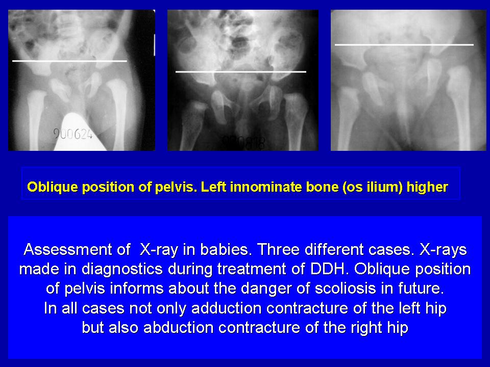
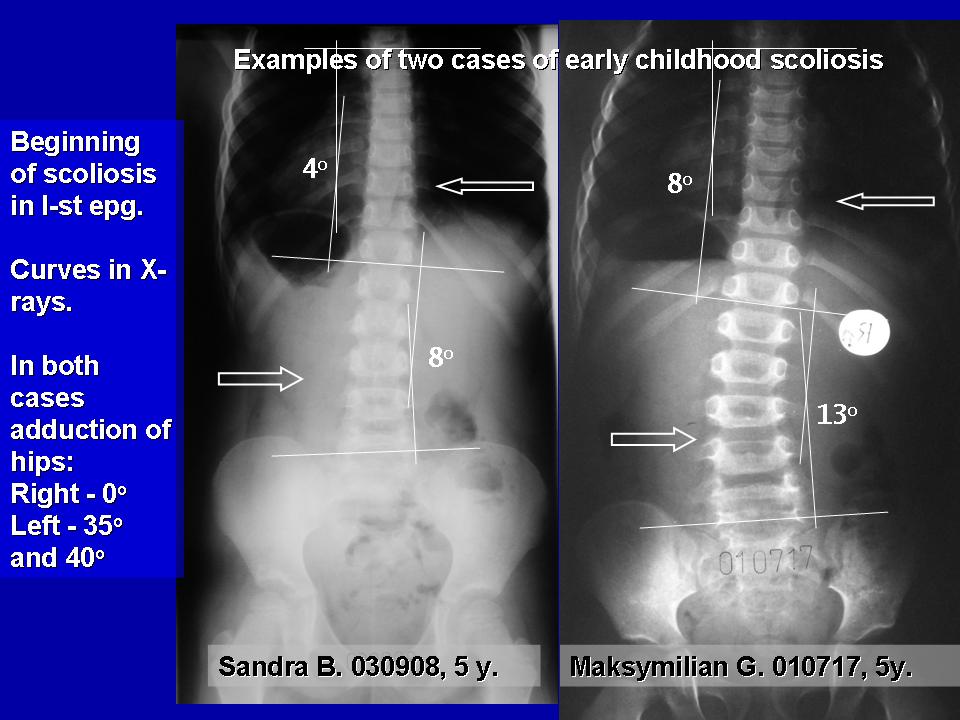
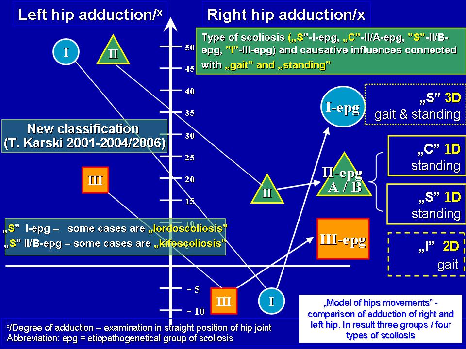
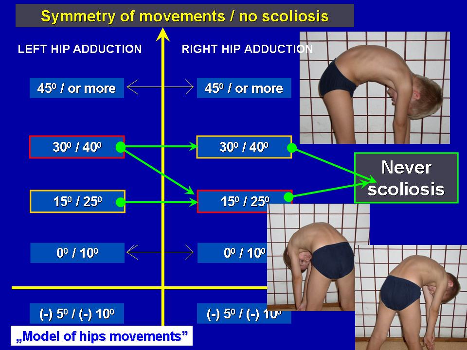
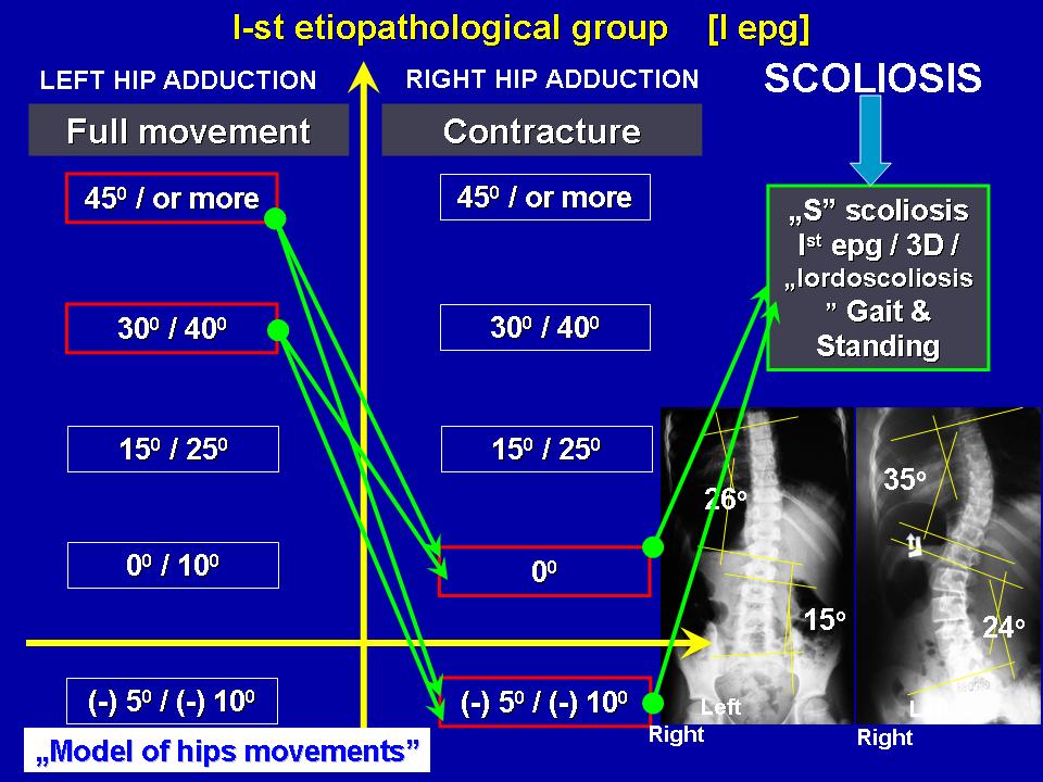
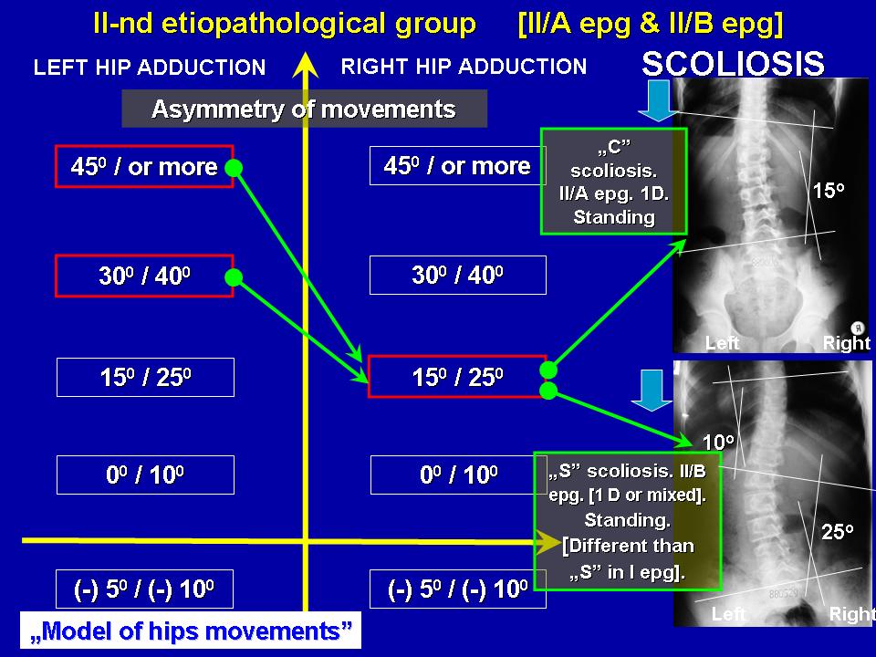
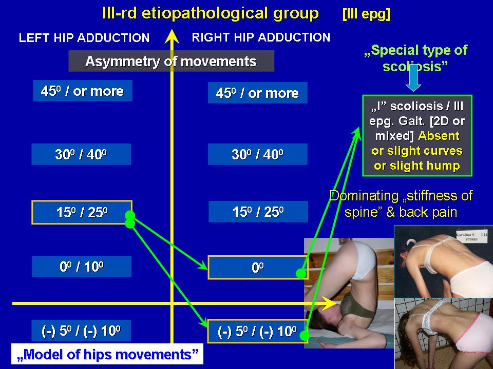
| Data modyfikacji: 2012-10-14 | The Web Side prepared by Prof. Tomasz Karski MD PhD. Lublin/Poland | |||
 |
 |